There are several aspects of the optical system which contribute to normal visual development in infants, and retinal development is essential for this. Retinal factors take 4+ years to reach full adult maturity. Receptors are found in the fovea and peripheral parts of the retina.
Vision rapidly improves after birth due to changes in the size, shape and density of cones found in the fovea. The outer segment length of the cones in the fovea is 16x shorter in newborns compared to adults. Visual acuity is further limited because the cone packing density is also 4x less in newborns which reduces spatial sampling. (VVP infant vision,2018)
Save your time!
We can take care of your essay
- Proper editing and formatting
- Free revision, title page, and bibliography
- Flexible prices and money-back guarantee
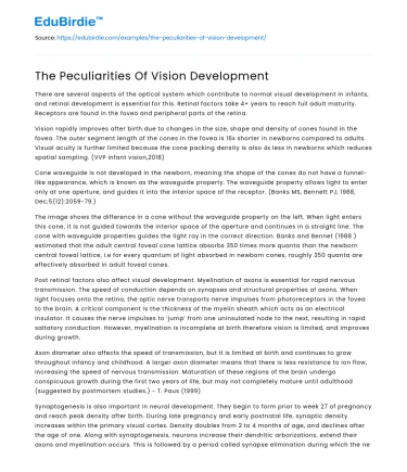
Cone waveguide is not developed in the newborn, meaning the shape of the cones do not have a funnel- like appearance, which is known as the waveguide property. The waveguide property allows light to enter only at one aperture, and guides it into the interior space of the receptor. (Banks MS, Bennett PJ, 1988, Dec;5(12):2059-79.)
The image shows the difference in a cone without the waveguide property on the left. When light enters this cone, it is not guided towards the interior space of the aperture and continues in a straight line. The cone with waveguide properties guides the light ray in the correct direction. Banks and Bennet (1988 ) estimated that the adult central foveal cone lattice absorbs 350 times more quanta than the newborn central foveal lattice, i.e for every quantum of light absorbed in newborn cones, roughly 350 quanta are effectively absorbed in adult foveal cones.
Post retinal factors also affect visual development. Myelination of axons is essential for rapid nervous transmission. The speed of conduction depends on synapses and structural properties of axons. When light focuses onto the retina, the optic nerve transports nerve impulses from photoreceptors in the fovea to the brain. A critical component is the thickness of the myelin sheath which acts as an electrical insulator. It causes the nerve impulses to ‘jump’ from one uninsulated node to the next, resulting in rapid saltatory conduction. However, myelination is incomplete at birth therefore vision is limited, and improves during growth.
Axon diameter also affects the speed of transmission, but it is limited at birth and continues to grow throughout infancy and childhood. A larger axon diameter means that there is less resistance to ion flow, increasing the speed of nervous transmission. Maturation of these regions of the brain undergo conspicuous growth during the first two years of life, but may not completely mature until adulthood (suggested by postmortem studies.) - T. Paus (1999)
Synaptogenesis is also important in neural development. They begin to form prior to week 27 of pregnancy and reach peak density after birth. During late pregnancy and early postnatal life, synaptic density increases within the primary visual cortex. Density doubles from 2 to 4 months of age, and declines after the age of one. Along with synaptogenesis, neurons increase their dendritic arborizations, extend their axons and myelination occurs. This is followed by a period called synapse elimination during which the nervous system fine tunes neural connectivity; which involves eliminating interconnections between redundant or non-functional neurones. This continues for over a decade. (VVP infant vision, 2018, slide 43)
To summarise, vision over the first 12 months experiences a rapid improvement. Although vision improves in the first few years of life, also known as the ‘slow juvenile phase’ the most rapid and critical changes occur during the rapid infantile phase due to emmetropization, retinal factors such as the development of cones and post retinal factors involving development of the visual cortex, myelination and synaptogenesis. These factors combined give rise to colour vision, improved contrast sensitivity and a higher visual acuity over the first 12 months of life.


 Stuck on your essay?

